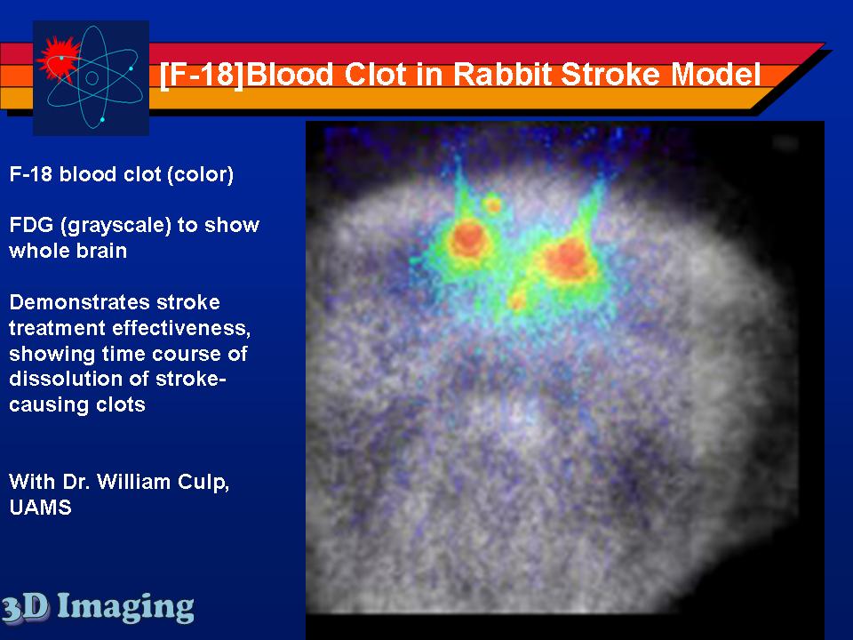
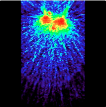
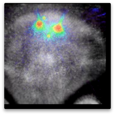
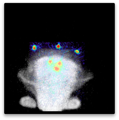
A natural stroke model in rabbits, in routine use in the laboratory Dr. Culp at UAMS, has been used for research into stroke progression and treatment. By addition of imaging to this model, the detailed progression of treatment of stroke has been measured. Clots formed from the rabbit's blood were radiolabeled by 3D Imaging with F-18 (I-124 and Zr-89 labeling is equally possible, as well as labeling for SPECT with I-125 or I-123, or even fluorescent labeling; but the use of PET allowed quantitative measurement of clot dissipation during a two hour period including treatment.). The radiolabeled blood clots were used by Dr. Culp's lab to create a natural stroke which was then treated. PET imaging before, during, and after treatment established that the clots were stable without treatment, degraded during treatment, and continued to degrade after treatment. These videos show the progression. In some only the PET scans are shown, so only the clots are visible, but are easily located and quantified. In others, an overlay of an FDG image is used to show the loacation of the brain and other soft tissues.
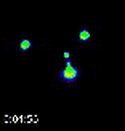
Videos show the field of view as it evolves in time, and with rotation to help visualize the 3D data and relative positions of the clots. Click on image to download and view the Videos.
Click on images to see additional examples, larger versions, and videos.